The Vertebrate Segmentation Clock: Converting Time into Embryonic Pattern
Presenter
March 7, 2008
Keywords:
- Cyclic homology
MSC:
- 19D55
Abstract
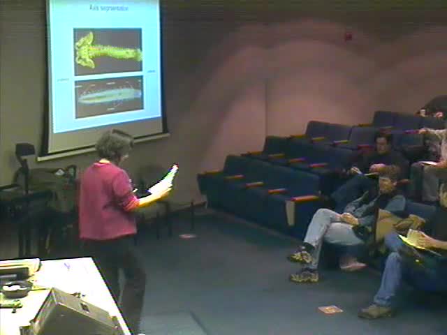
The vertebrate body can be subdivided along the antero-posterior (AP) axis into repeated structures called segments. This periodic pattern is established during embryogenesis by the somitogenesis process. Somites are generated in a rhythmic fashion from the paraxial mesoderm and subsequently differentiate to give rise to the vertebrae and skeletal muscles of the body. Somite formation involves an oscillator, the segmentation clock whose periodic signal is converted into the periodic array of somite boundaries. This clock drives the dynamic expression of cyclic genes in the presomitic mesoderm and requires Notch and Wnt signaling. Microarray studies of the mouse presomitic mesoderm transcriptome reveal that the segmentation clock drives the periodic expression of a large network of cyclic genes involved in cell signaling. Mutually exclusive activation of the Notch/FGF and Wnt pathways during each cycle, suggests that coordinated regulation of these three pathways underlies the clock oscillator. Whereas the segmentation clock is thought to set the pace of vertebrate segmentation, the translation of this pulsation into the reiterated arrangement of segment boundaries along the AP axis involves FGF and Wnt signaling. The FGF pathway controls the positioning of the wavefront, which corresponds to the level of the presomitic mesoderm where cells respond to the clock. fgf8 mRNA is only transcribed in tail bud precursors and it progressively decays in newly formed paraxial mesoderm cells, thus forming a dynamic mRNA gradient. This mRNA gradient is then translated into a graded FGF signaling response used to position the wavefront. This mechanism provides an efficient means to couple the spatio-temporal activation of segmentation to the posterior elongation of the embryo.