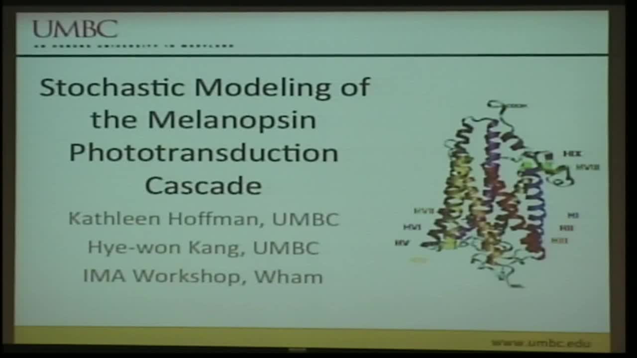
Stochastic Modeling of the Phototransduction Cascade for Melanopsin
Presenters
September 9, 2013
Abstract
Phototransduction cascades transform light to electrical signals that project to the brain through a sequence of chemical reactions. Image forming vertebrate vision is facilitated by rods and cones in the back of the retina, each of which has its own opsin, a light sensitive G-protein coupled receptor. Recently a third opsin, melanopsin, was identified, not in rods and cones, but in intrinsically photosensitive retinal ganglion cells (ipRGCs). It is thought that melanopsin is primarily responsible for non-‐image forming visual functions such as the regulation of sleep cycles and pupillary light reflex.
Since melanopsin was only discovered recently, not much is known about its phototransduction pathway, which involves both an activation and inactivation pathway, however, it is believed that melanopsin uses a pathway similar to those found in invertebrate retinas. Data suggests that the melanopsin pathway consists of an activated G-‐protein, which in turn activates a phospholipase‐C (PLC). PLC breaks down PIP2 into second messengers, which leads to an increase in the intracellular calcium, and in turn depolarizes the cell. Inactivation of the melanopsin is hypothesized to be a two part process that first involves phosphorylation of its carboxy tail and second binding by a beta-‐arrestin because melanopsin is a G-‐protein coupled receptor.
Based on modeling of rhodopsin, a well-known and well-‐studied rod photopigment, a differential equation based model of the activation and inactivation of melanopsin has been formulated and tested. Both the activation and the inactivation can be described by a phototransduction cascade, which consists of a series of chemical reactions. For both activation and inactivation, the model has been tested against two types of experimental data: electrophysiology data and calcium imaging data from melanopsin cultivated in human embryonic kidney (HEK) cells. Model parameters that weren’t obtained from experimental data were fit using the wild type data. The model of inactivation, with the parameters from the wild type data, accurately predicted experimental responses to an over expression of beta arrestin. Current work involves coupling the two models to predict the response to a second flash of light that experimentally produces a quicker response than the first flash, yet with lower amplitude, a process known as adaptation.
Hamer et al (2003) developed a model for rhodopsin phototransduction cascade that included stochastic mechanisms in which the probability that a molecular species engages in a particular reaction is proportional to the reaction rate and then use Monte-Carlo simulations and the Gillespie method to accurately predict activation and inactivation of rhodopsin. In this project, I propose to extend the methods suggested by Hamer et al for rhodopsin to melanopsin.
Hamer, Nicholas, Tranchina, Leiderman, Lamb, Multiple Steps of Phosphorylation of Activated Rhodopsin can account for the Reproducibility of Vertebrate Rod Single-photon Responses, J. Gen. Physiology, vol 122, 419‐444, 2003.
